
Case 7
Dr Marina-Portia Anthony /Dr Henry Mak /Dr Jingbo Zhang
Clinical notes
A 59 year-old female complained of intermittent postmenopausal bleeding. An ultrasound showed thickened endometrium, and a biopsy revealed endometrioid endometrial adenocarcinoma. A PET-CT was requested for staging.Images
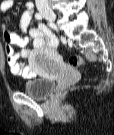
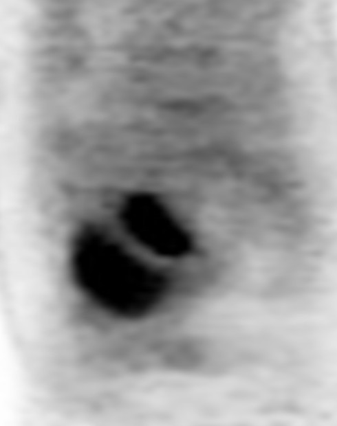
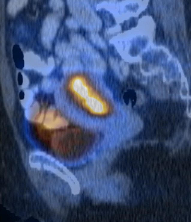
Figure 1.
Reformatted sagittal contrast-enhanced CT (A), FDG-PET (B) and fused PET-CT (C) images of the pelvis.
Show the video
References
- Iyer R. Balachandran A. Devine C. PET/CT and cross sectional imaging of gynecologic malignancy. Cancer Imaging. 2007; 7(A): S130-8.
- Lee S. Russell A. Performance of 18F-FDG PET/CT in endometrial cancer. AJR Am J Roentgenol. 2008; 191(5): W210.
- Lerman H. Metser U. Grisaru D. et al. Normal and abnormal 18F-FDG endometrial and ovarian uptake in pre- and postmenopausal patients: assessment by PET/CT. J Nucl Med. 2004; 45(2): 266-71.
- Lin EC, Alavi A. PET and PET/CT. New York, Thieme Medical Publishers Inc., 2005:45.
- Park J. Kim E. Kim D. et al. Comparison of the validity of magnetic resonance imaging and positron emission tomography/computed tomography in the preoperative evaluation of patients with uterine corpus cancer. Gynecol Oncol. 2008; 108(3): 486-92.
- Kumar V. Abbas A. Fausto N. Robbins and Cotran Pathologic Basis of Disease. 2005. Elselvier. Pennsylvania.