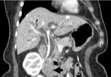
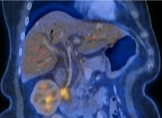
Adenocarcinoma of the ampulla of Vater is relatively uncommon, accounting for approximately 0.2% of gastrointestinal tract malignancies and approximately 7% of all periampullary carcinomas. Ampullary carcinomas can be located in the ampulloduodenal part of the ampulla of Vater lined by intestinal mucosa, or in the deeper part of the ampulla lined by pancreaticobiliary duct mucosa. Thus the two main histologic subtypes are the intestinal-type adenocarcinoma and pancreaticobiliary-type adenocarcinoma. In many cases, symptoms due to stenosis lead to diagnosis at an early tumor stage. In about 80%, curative resection is possible.
18-FDG-PET is very sensitive for detecting periampullary neoplasms. It may identify invasive tumors even when they are difficult to visualize on standard imaging. It is also useful for follow-up of post-resection patients to identify recurrence.


