Case 57
Dr Marina-Portia Anthony / Professor Pek-Lan Khong
Clinical notes
A 4 year-old boy presented for further investigation of abdominal discomfort.Images
Figure 1.
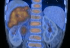
Reformatted coronal contrast-enhanced CT (A) and fused FDG-PET/CT (B) images through the chest and abdomen.
Figure 2.
Reformatted coronal contrast-enhanced CT (A) and fused FDG-PET/CT (B) images through the upper abdomen.
Show the video
References
- Dahnert W. Radiology Review Manual. 2003. Lippincott Williams & Wilkins. Philadelphia.
- Kumar V. Abbas A. Fausto N. Robbins and Cotran Pathologic Basis of Disease. 2005. Elselvier. Pennsylvania.
- Kushner BH, et al. Extending positron emission tomography scan utility to high-risk neuroblastoma: fluorine-18 fluorodeoxyglucose positron emission tomography as sole imaging modality in follow-up of patients. J Clin Oncol 2001;19:3397-405.
- Shulkin BL, Hutchinson RJ, Castle VP, Yanik GA, Shapiro B, Sisson JC. Neuroblastoma: positron emission tomography with 2-[fluorine-18]-fluoro-2-deoxy-D-glucose compared with metaiodobenzylguanidine scintigraphy. Radiology. 1996;199(3):743-50.


