Dr Marina-Portia Anthony / Professor Pek-Lan Khong
Clinical notes
An 11-year-old girl presented with a 3 month history of right knee pain and swelling. There was no history of trauma or fever.
Images
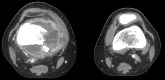
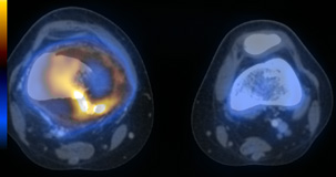
Figure 1.
Axial contrast-enhanced CT (A) and fused FDG-PET/CT (B) images through the distal femora.
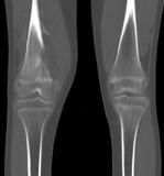
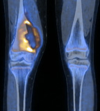
Figure 2.
Reformatted coronal contrast-enhanced CT on bone algorithm (A) and fused FDG-PET/CT (B) images of the distal femora.
Show the video
Findings
Figures 1,2. In the right medial distal femoral metaphysis, there is a hypermetabolic lytic lesion (SUVmax 5.1) with a large soft tissue component. The FDG uptake is predominantly around the rim of the lesion, with central areas of low metabolic activity present. There is periosteal reaction and mild bone sclerosis. Distally, the lesion extends across the physeal plate to the proximal epiphysis. Medially, there is cortical disruption. Posteriorly, the popliteal vessels are displaced.
Figure 3. No other hypermetabolic lesions are present.
Discussion
Osteosarcomas most commonly occur in the femur, with the metaphysis being the commonest location within long bones. Fused FDG-PET/CT significantly improves differentiation between malignant and benign bone lesions compared to PET alone. In addition, FDG uptake has been shown to correlate with tumour grade, and this information has also been found to be of prognostic significance. Delayed imaging may also help to differentiate malignant from benign lesions, with malignant lesions showing increased FDG uptake on delayed images.
False negative interpretation can occur in the presence of necrosis (low metabolic activity, as seen in the centre of the lesion in the current case), and also in some other types of malignant primary bone tumour, including low-grade chondrosarcoma, plasmacytoma and myxoid tumours.
References
- Adler LP, Blair HF, Makley JT, Williams RP, Joyce MJ, Leisure G, et al. Non-invasive grading of musculoskeletal tumours using PET. J Nucl Med. 1991 Aug;32(8):1508-12.
- Franzius C, Sciuk J, Daldrup-Link HE, Jürgens H, Schober O. FDG-PET for detection of osseous metastases from malignant primary bone tumours: comparison with bone scintigraphy. Eur J Nucl Med. 2000;27(9):1305-11.
- Lin EC, Alavi A. PET and PET/CT. 2005. Thieme Medical Publishers Inc. New York.
- Strobel K, Exner UE, Stumpe KD, Hany TF, Bode B, Mende K, et al. The additional value of CT images interpretation in the differential diagnosis of benign vs. malignant primary bone lesions with 18F-FDG-PET/CT. Eur J Nucl Med Mol Imaging. 2008;35(11):2000-8.
- Von Shulthess G. Molecular Anatomic Imaging. 2007.Lippincott Williams & Willkins. Philadelphia.


