Case 54
Dr Marina-Portia Anthony / Professor Pek-Lan Khong
Clinical notes
A 13-year-old male presented for a follow-up scan to assess treatment response of a left maxillary sinus tumour.Images
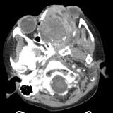
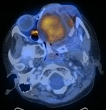
Figure 1.
Axial contrast-enhanced CT (A) and fused FDG-PET/CT (B) images through the mid-face.
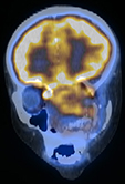
Figure 2.
Reformatted coronal contrast-enhanced CT (A) and fused FDG-PET/CT (B) images of the mid-face.
Show the video
References
- Kumar V. Abbas A. Fausto N. Robbins and Cotran Pathologic Basis of Disease. 2005. Elselvier. Pennsylvania.
- McCarville MB, Christie R, Daw NC, Spunt SL, Kaste SC. PET/CT in the evaluation of childhood sarcomas. AJR Am J Roentgenol. 2005;184(4):1293-304.
- Peng F, Rabkin G, Muzik O. Use of 2-deoxy-2-[F-18]-fluoro-D-glucose positron emission tomography to monitor therapeutic response by rhabdomyosarcoma in children: report of a retrospective case study. Clin Nucl Med. 2006;31(7):394-7.
- Tateishi U, Hosono A, Makimoto A, Nakamoto Y, Kaneta T, Fukuda H, et al. Comparative study of FDG PET/CT and conventional imaging in the staging of rhabdomyosarcoma. Ann Nucl Med. 2009;23(2):155-61.
- Völker T, Denecke T, Steffen I, Misch D, Sch?nberger S, Plotkin M, et al. Positron emission tomography for staging of pediatric sarcoma patients: results of a prospective multicenter trial. J Clin Oncol. 2007;25(34):5435-41.


