Dr Marina-Portia Anthony / Professor Pek-Lan Khong
Clinical notes
A 1-year-old male presented for liver transplantation work-up, on a background of known Stage III multifocal hepatoblastoma.
Images
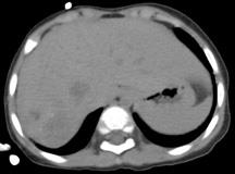
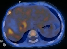
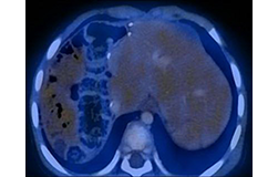
Figure 1.
Axial unenhanced CT scan (A) and fused FDG-PET/CT (B) images through the upper abdomen.
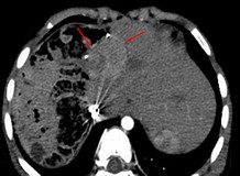
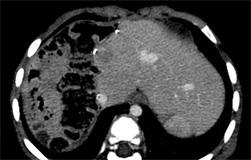
Figure 2.
Axial contrast-enhanced CT scan of another patient with suspected hepatoblastoma recurrence, post surgery, in the arterial phase (A), porto-venous phase (B), and fused FDG-PET/CT images (C).
Findings
Figure 1. There is a hypodense mass lesion in the right lobe of the liver with several foci of low-grade metabolic activity (up to SUVmax 1.6, with background liver activity of SUVmax 0.9). On arterial phase imaging (not shown), multiple arterial enhancing foci were seen, with subsequent washout.
Figure 2. Arterial phase contrast-enhanced CT shows a hypervascular, arterial enhancing tumour at the resection margin and wash-out in the porto-venous phase (B). FDG-PET/CT did not show increased uptake (C).
Discussion
Hepatoblastoma is the commonest primary liver tumour in children, and the third commonest abdominal tumour in children. Accurate localization is important for biopsy purposes, and for appropriate management. The alpha-foetoprotein is frequently markedly raised, and also increases in cases of recurrence. The role of FDG PET/CT is not well-established due to limited experience. Anecdotal experience has found that tumours to have variable FDG-avidity.
References
- Dahnert W. Radiology Review Manual. 2003. Lippincott Williams & Wilkins. Philadelphia.
- Figarola MS, McQuiston SA, Wilson F, Powell R. Recurrent hepatoblastoma with localization by PET-CT. Pediatr Radiol. 2005;35(12):1254-8.
- Philip I, Shun A, McCowage G, Howman-Giles R. Positron emission tomography in recurrent hepatoblastoma. Pediatr Surg Int. 2005;21(5):341-5.


