Dr Marina-Portia Anthony
Clinical notes
A 52 year-old female presented with recent increase in size of her known diffuse goitre of 6 years.
Images
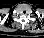
Figure 1.
Axial (A) and reformatted coronal (B) contrast-enhanced CT images of the neck.
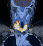
Figure 2.
Axial (A) and reformatted coronal (B) fused FDG-PET/CT images of the neck.
Show the video
Findings
Figures 1,2. Both lobes of the thyroid glands are markedly enlarged with homogeneous enhancement and markedly increased metabolic activity (SUVmax 10.9).
Figure 3. In addition to the hypermetabolic activity seen in the enlarged thyroid gland, there is a hypermetabolic (SUVmax 3.8) lymph node in the left para-tracheal region, and a mildly hypermetabolic (SUVmax 2.0) left level II lymph node in the neck.
Diagnosis
MALT lymphoma of the thyroid gland. No co-existing thyroiditis was reported histopathologically.
Discussion
The normal thyroid has a mean standardized uptake value of 1.3. Mild diffuse FDG uptake in the thyroid gland may be a normal variant, however greater degrees are usually pathologic. Whilst FDG uptake in diffuse large B-cell lymphoma of the thyroid has been reported, few reports about thyroid MALT lymphoma with FDG uptake are published. Because of the frequent coexistence of chronic thyroiditis and MALT lymphoma, no difference in the degree of hypermetabolism may be demonstrated. Thus FDG-PET imaging may not be indicated to evaluate the status of the MALT lymphoma in these patients.
References
- Lee CJ, Hsu CH, Tai CJ, et al. FDG-PET for a thyroid MALT lymphoma. Acta Oncol. 2008;47(6):1165-7.
- Lin EC, Alavi A. PET and PET/CT. 2005. Thieme Medical Publishers Inc. New York.
- Mikosch P, Würtz FG, Gallowitsch HJ, et al. F-18-FDG-PET in a patient with Hashimoto's thyroiditis and MALT lymphoma recurrence of the thyroid. Wien Med Wochenschr. 2003;153(3-4):89-92.


