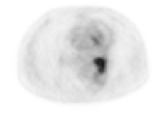
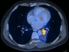
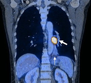
FDG-PET-CT is a valuable tool for evaluating the nature of a pulmonary nodule, though one must be aware of causes of false positives and negatives. A suspicious nodule with an SUVmax 2.5 or higher has a high chance of representing malignancy (up to 75% in some studies). PET-CT may also help differentiate the histologic subtype of lung tumour in some cases, using combined information including tumor size, CT density, and metabolic activity. For example, bronchioalveolar cell carcinoma tends to have a smaller size, lower CT density, and lower FDG avidity compared to adenocarcinoma.
This case shows that PET-CT is useful for evaluating primary tumour extent, and distinguishing tumor from benign post-obstructive change. PET-CT is also a valuable diagnostic tool for initial systemic staging of lung cancer. Repeat PET imaging after the initiation of chemoradiotherapy can predict tumor response and help tailor therapy. The European Organization for Research and Treatment of Cancer (EORTC) criterion for partial response on FDG-PET is 25% reduction in SUVmax.


