Dr Marina-Portia Anthony
Clinical notes
A 35 year-old female presented for staging after excision of a left thigh synovial sarcoma. She had recently experienced left calf numbness.
Images
Figure 1.
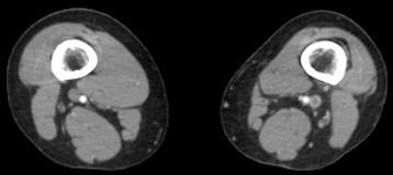
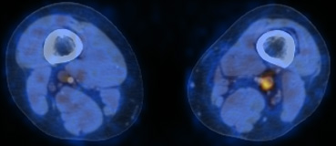
Axial contrast-enhanced CT (A) and fused FDG-PET/CT (B) images of the lower thighs.
Findings
There is a filling defect and hypermetabolic activity in the enlarged left popliteal vein.
Diagnosis
Deep vein thrombosis. This was confirmed sonographically.
Discussion
Venous thrombosis is known to incite an inflammatory response within the vein lumen and vessel wall, mediated by cytokines and leukocytes. This is well demonstrated on PET/CT, even in the absence of intravenous contrast, with FDG uptake seen in venous structures with any of: bland (non-infected) thrombi, tumour thrombosis, and septic thrombophlebitis. Uptake may occur in acute or chronic thrombosis. This inflammatory response has been corroborated by MRI, where gadolinium enhancement of the vessel wall has been demonstrated. The FDG uptake in venous thrombosis may simulate a neoplastic process, and is thus an important pitfall in FDG-PET/CT imaging.
References
- Do B, Mari C, Biswal S, et al. Diagnosis of aseptic deep venous thrombosis of the upper extremity in a cancer patient using fluorine-18 fluorodeoxyglucose positron emission tomography/computerized tomography (FDG PET/CT). Ann Nucl Med. 2006;20(2):151-5.
- Lin EC, Alavi A. PET and PET/CT. 2005. Thieme Medical Publishers Inc. New York.
- Ryan M. Deep venous thrombosis in the deep posterior compartment of the lower limb: PET CT FDG findings. Clin Nucl Med. 2008;33(8):582-4.
- Sydow BD, Srinivas SM, Newberg A, Alavi A. Deep venous thrombosis on F-18 FDG PET/CT imaging. Clin Nucl Med. 2006;31(7):403-4.


