
Case 48
Dr Marina-Portia Anthony
Clinical notes
A 41 year-old female presented with left knee pain. A plain radiograph showed an osteolytic lesion in the distal left femur.Images
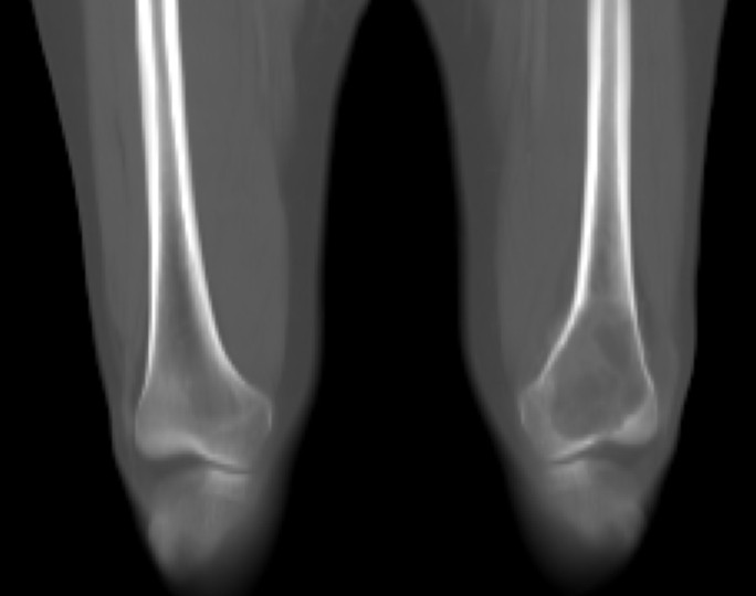
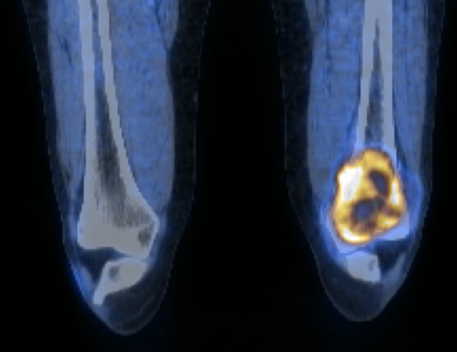
Figure 1.
Reformatted coronal contrast-enhanced CT (A) and fused FDG-PET/CT (B) images of the lower femora.
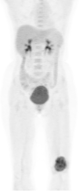
Figure 2.
Coronal MIP FDG-PET
References
- Aoki J, Watanabe H, Shinozaki T, et al. FDG PET of primary benign and malignant bone tumors: standardized uptake value in 52 lesions. Radiology. 2001;219(3):774-7.
- Kumar V. Abbas A. Fausto N. Robbins and Cotran Pathologic Basis of Disease. 2005. Elselvier. Pennsylvania.
- Lin EC, Alavi A. PET and PET/CT. 2005. Thieme Medical Publishers Inc. New York.
- McKinney AM, Reichert P, Short J, et al. Metachronous, multicentric giant cell tumor of the sphenoid bone with histologic, CT, MR imaging, and positron-emission tomography/CT correlation. AJNR Am J Neuroradiol. 2006;27(10):2199-201.