Case 46
Dr Marina-Portia Anthony
Clinical notes
A 73 year-old man presented with an episode of haematemesis and dysphagia to solid foods for 3 months. He had a history of long term alcohol use and smoking.Images
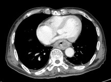
Figure 1.
Axial (A) and reformatted sagittal (B) contrast-enhanced CT images of the lower chest.
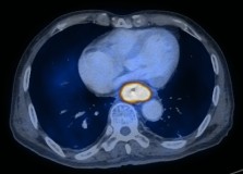
Figure 2.
Axial (A) and reformatted sagittal (B) fused FDG-PET/CT images of the lower chest.
References
- Guo H, Zhu H, Xi Y, Zhang B, et al. Diagnostic and prognostic value of 18F-FDG PET/CT for patients with suspected recurrence from squamous cell carcinoma of the esophagus. J Nucl Med. 2007;48(8):1251-8.
- Kumar V. Abbas A. Fausto N. Robbins and Cotran Pathologic Basis of Disease. 2005. Elselvier. Pennsylvania.
- Lin EC, Alavi A. PET and PET/CT. 2005. Thieme Medical Publishers Inc. New York.
- Roedl JB, Colen RR, King K, et al. Visual PET/CT scoring for nonspecific 18F-FDG uptake in the differentiation of early malignant and benign esophageal lesions. AJR Am J Roentgenol. 2008;191(2):515-21.
- Roedl JB, Halpern EF, Colen RR, et al. Metabolic tumor width parameters as determined on PET/CT predict disease-free survival and treatment response in squamous cell carcinoma of the esophagus. Mol Imaging Biol. 2009;11(1):54-60.


