Case 42
Dr Marina-Portia Anthony /Dr Tao Chan
Clinical notes
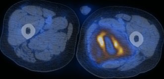
A 70 year-old man with a background of myelofibrosis presented with a persistent low-grade fever. Septic workup was negative. An FDG-PET/CT scan was requested to localize potential foci of infection.Images
Figure 1.
Axial unenhanced CT (A), FDG-PET (B) and fused FDG-PET/CT (C) images through the upper thighs.
Show the video
References
- Castellucci P, Nanni C, Farsad M, Alinari L, Zinzani P, Stefoni V, et al. Potential pitfalls of 18F-FDG PET in a large series of patients treated for malignant lymphoma: prevalence and scan interpretation. Nucl Med Commun. 2005;26(8):689-94.
- Lin EC, Alavi A. PET and PET/CT. 2005. Thieme Medical Publishers Inc. New York.
- Metser U, Miller E, Lerman H, Even-Sapir E. Benign nonphysiologic lesions with increased 18F-FDG uptake on PET/CT: characterization and incidence. AJR Am J Roentgenol. 2007;189(5):1203-10.
- Sugawara Y, Braun DK, Kison PV, Russo JE, Zasadny KR, Wahl RL. Rapid detection of human infections with fluorine-18 fluorodeoxyglucose and positron emission tomography: preliminary results. Eur J Nucl Med. 1998;25(9):1238-43.
- Von Shulthess G. Molecular Anatomic Imaging. 2007.Lippincott Williams & Willkins. Philadelphia.


