Case 40
Dr Marina-Portia Anthony /Dr Tao Chan
Clinical notes
A 44 year-old woman presented with abdominal distension and diarrhoea for 1 month, and a past history of right dermoid cyst with cystectomy.Images
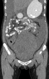
Figure 1.
Reformatted coronal unenhanced CT (A) and fused FDG-PET/CT (B) images of the pelvis.
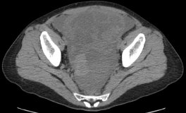
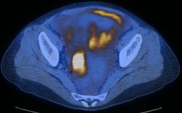
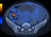
Figure 2.
Axial unenhanced CT (A) and fused FDG-PET/CT (B) images of the pelvis.
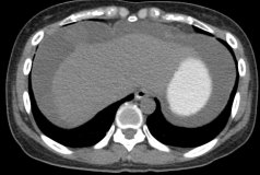
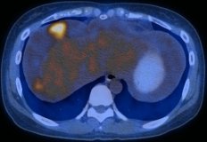
Figure 3.
Axial contrast-enhanced CT (A) and fused FDG-PET/CT (B) images of the pelvis.
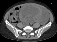
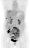
Figure 4.
Axial contrast-enhanced CT (A) and fused FDG-PET/CT (B) images of the pelvis.
Show the video
References
- Anthony MP, Khong PL, Zhang JB. Spectrum of 18F-FDG-PET/CT appearances in peritoneal disease. Am J Roentgenol 2009;193:W523-9.
- Bohdiewicz PJ, Juni JE, Ball D, et al. Krukenberg tumor and lung metastases from colon carcinoma diagnosed with F-18 FDG PET. Clin Nucl Med. 1995 May;20(5):419-20.
- Dahnert W. Radiology Review Manual. 2003. Lippincott Williams & Wilkins. Philadelphia.
- Henley T, Reddy MP, Ramaswamy MR, et al. Bilateral ovarian metastases from colon carcinoma visualized on F-18 FDG PET scan. Clin Nucl Med. 2004 May;29(5):322-3.
- Kumar V. Abbas A. Fausto N. Robbins and Cotran Pathologic Basis of Disease. 2005. Elselvier. Pennsylvania.
- Lin EC, Alavi A. PET and PET/CT. 2005. Thieme Medical Publishers Inc. New York.
- Von Shulthess G. Molecular Anatomic Imaging. 2007.Lippincott Williams & Willkins. Philadelphia.


