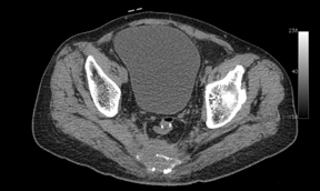
The distribution of carcinoma in the large intestine is as follows: rectosigmoid colon 55%, caecum/ ascending colon 22%, transverse colon 11%, descending colon 6%, and other sites 6%. Whilst all colorectal carcinomas begin as in situ lesions, they evolve into different morphologic patterns, with distal tumours tending to be annular, encircling lesions with proximal heaped margins and ulcerated mid-regions. This causes luminal narrowing and proximal bowel distension or obstruction.
Conventional imaging methods for rectal carcinoma, such as barium enema, CT and MRI, are helpful in defining initial tumour extent at diagnosis, but are not accurate in distinguishing residual or recurrent tumour from scar tissue after chemotherapy and/ or surgical resection, which is important for surgical planning. FDG-PET-CT has been shown to have both high sensitivity and high specificity in the detection of local disease recurrence.


