Case 38
Dr Marina-Portia Anthony /Dr Tao Chan
Clinical notes
A 1 year-old male presented with prolonged fever and lymph node enlargement after liver transplantation for biliary cirrhosis.Images
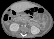
Figure 1.
Axial contrast-enhanced CT (A) and fused FDG-PET/CT images (B) of the upper abdomen.
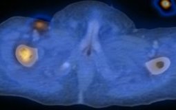
Figure 2.
Axial contrast-enhanced CT (A) and fused FDG-PET/CT images (B) through the inguinal regions.
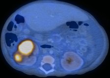
Figure 3.
Axial unenhanced CT (A) and fused FDG-PET/CT images (B) through the kidneys.
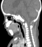
Figure 4.
Reformatted sagittal contrast-enhanced CT (A) and fused FDG-PET/CT images (B) through the nasopharynx.
Show the video
References
- von Falck C, Maecker B, Schirg E, et al. Post transplant lymphoproliferative disease in pediatric solid organ transplant patients: a possible role for [18F]-FDG-PET(/CT) in initial staging and therapy monitoring. Eur J Radiol. 2007;63(3):427-35.
- Bakker NA, van Imhoff GW, Verschuuren EA, et al. Presentation and early detection of post-transplant lymphoproliferative disorder after solid organ transplantation. Transpl Int. 2007;20(3):207-18.
- McCormack L, Hany TI, H?bner M, et al. How useful is PET/CT imaging in the management of post-transplant lymphoproliferative disease after liver transplantation? Am J Transplant. 2006;6(7):1731-6.


