Case 36
Dr Marina-Portia Anthony /Dr Tao Chan
Clinical notes
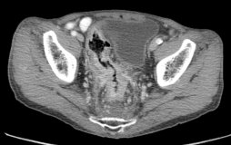
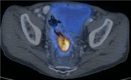
A 76 year-old male complained of bright rectal bleeding over the previous 1 year, and altered bowel habit with weight loss. Per rectal examination revealed a circumferential mass at tip of finger.Images
Figure 1.
Axial contrast-enhanced CT (A) and fused PET/CT (B) images through the lower pelvis
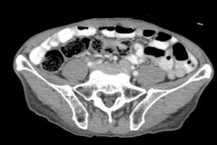
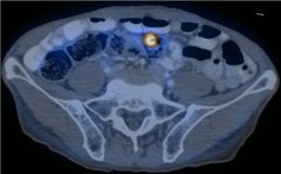
Figure 2.
Axial contrast-enhanced CT (A) and fused PET/CT (B) images through the upper pelvis.
Show the video
References
- Kamel I, Cohade C, Neyman E. et al. Incremental value of CT in PET/CT of patients with colorectal carcinoma. Abdom Imaging. 2004;29(6):663-8.
- Kumar V. Abbas A. Fausto N. Robbins and Cotran Pathologic Basis of Disease. 2005. Elselvier. Pennsylvania.
- Mainenti P, Salvatore B, D'Antonio D. et al. PET/CT colonography in patients with colorectal polyps: a feasibility study. Eur J Nucl Med Mol Imaging. 2007;34(10):1594-603.
- Park I, Kim H, Yu C, at al. Efficacy of PET/CT in the accurate evaluation of primary colorectal carcinoma. Eur J Surg Oncol. 2006;32(9):941-7.


