
Case 30
Dr Marina-Portia Anthony /Dr Tao Chan
Clinical notes
A 64 year old male presented with fever, without localising signs. The patient had past history of autoimmune hemolytic anaemia, splenectomy and portal vein thrombosis. A CT chest and abdomen showed multiple pulmonary and liver nodules. A PET-CT was requested to look for a source of infection or malignancy.Images
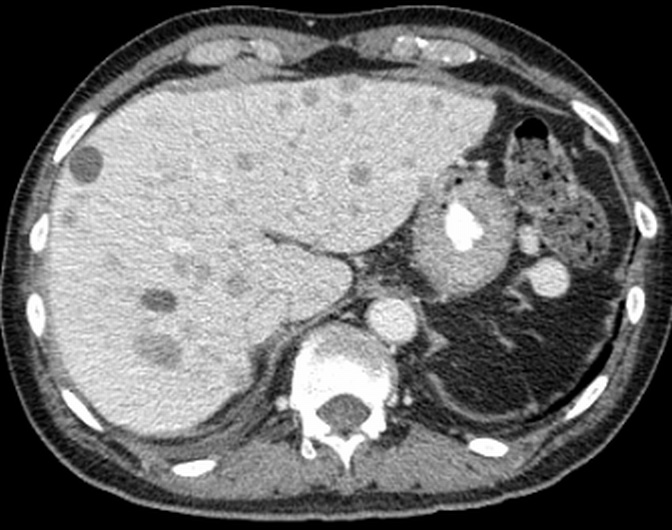
A.
CE axial CT abdomen
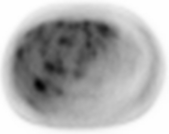
B.
Axial FDG-PET abdomen
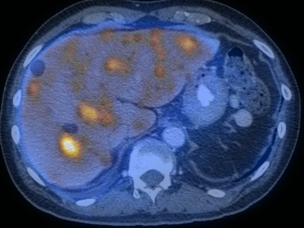
C.
Axial fused PET-CT abdomen
Show the video
References
- Chan WKS, Tse EWC, Fan PYS et al. Positron emission tomography/computed tomography in the diagnosis of multifocal primary hepatic lymphoma. J Clin Oncol 2008;26:5479-80.
- Kasamon Y. Jones R. Wahl R. Integrating PET and PET/CT into the risk-adapted therapy of lymphoma. J Nucl Med. 2007; 48(suppl): 19S-27S.
- Kumar V. Abbas A. Fausto N. Robbins and Cotran Pathologic Basis of Disease. 2005. Elselvier. Pennsylvania.
- Pelosi E. Pregno P. Penna D. et al. Role of whole-body [18F] fluorodeoxyglucose positron emission tomography/computed tomography (FDG-PET/CT) and conventional techniques in the staging of patients with Hodgkin and aggressive non Hodgkin lymphoma. Radiol Med. 2008; 113(4): 578-90.
- Specht L. 2-[18F]fluoro-2-deoxyglucose positron-emission tomography in staging, response evaluation, and treatment planning of lymphomas. Semin Radiat Oncol. 2007; 17(3): 190-7.