Case 3
Dr Marina-Portia Anthony / Dr Alex Ching /Dr Jingbo Zhang
Clinical notes
A 72 year-old male presented for post-treatment evaluation of his known multiple myeloma with extramedullary disease.Images
Figure 1.
Axial contrast-enhanced CT (A) and fused PET/CT (B) images through the upper chest.
Figure 2.
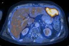
Axial contrast-enhanced CT (A) and fused PET/CT (B) images through the upper abdomen.
PLease add Data Source
PLease add Data Source
Figure 3.
Axial contrast-enhanced CT (A) and fused PET/CT (B) images through the lower abdomen.
Show the video
References
- Dankerl A, Liebisch P, Glatting G, et al. Multiple Myeloma: Molecular Imaging with 11C-Methionine PET/CT--Initial Experience. Radiology. 2007;242(2):498-508.
- Kumar V. Abbas A. Fausto N. Robbins and Cotran Pathologic Basis of Disease. 2005. Elselvier. Pennsylvania.
- Mulligan ME, Badros AZ. PET/CT and MR imaging in myeloma. Skeletal Radiol. 2007 Jan;36(1):5-16.
- Wiesenthal AA, Nguyen BD. F-18 FDG PET/CT staging of multiple myeloma with diffuse osseous and extramedullary lesions. Clin Nucl Med. 2007;32(10):797-801.


