Dr Marina-Portia Anthony /Dr Henry Mak
Clinical notes
A 22 year-old woman with a history of Hodgkin's lymphoma stage IIb, presented for a post-treatment FDG-PET/CT scan.
Images
Figure 1.
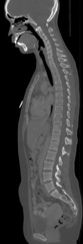
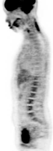
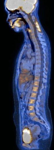
Reformatted sagittal CT (A), FDG-PET (B) and fused FDG-PET/CT (C) images of the spine.
Findings
Figure 1. There is osteopaenia and marked reduction in normal metabolic activity in the upper and mid- thoracic vertebrae.
Discussion
Reduced metabolic activity within the marrow of irradiated vertebrae (or vertebrae within radiotherapy portals) is revealed with FDG-PET/CT. This is attributed to fatty replacement of the marrow, with fewer residual capillaries; and vasculitis and hyalinization of small vessels in the vertebrae, with interruption of the blood supply, the combination of which leads to photopaenia. Other causes of reduced FDG activity in vertebrae include: some metastatic disease, and vertebral haemangioma amid increased marrow uptake after colony-stimulating factor therapy (relative reduction).
References
- Basu S, Nair N. "Cold" vertebrae on F-18 FDG PET: Causes and characteristics. Clin Nucl Med. 2006;31(8):445-50.
- Blau M, Gonatra R, Bender MA. F-18 FDG for bone imaging. Semin Nucl Med 1972; 2:31-7.
- King MA, Casarett GW, Weber DA. A study of irradiated bone: I. Histologic and physiologic changes. J Nucl Med 1979; 20:1142-9.
- Knope WH, Blom J, Grosby WH. Regeneration of locally irradiated bone marrow. I. Dose dependent, long-term changes in the rat with particular emphasis upon vascular and stoma reaction. Blood 1966; 28:398-415.
- Meyer MA, Nathan CO. Reduced F-18 fluorodeoxyglucose uptake within marrow after external beam radiation. Clin Nucl Med. 2000;25(4):279-80.
- Shih WJ. Incidental detection of diminished bone marrow metabolic activity with FDG PET. Radiographics. 2001;21(2):519.


