Case 27
Dr Marina-Portia Anthony /Dr Henry Mak
Clinical notes
A 26-year old man presented with fever, on a background of systemic lupus erythematosis.Images
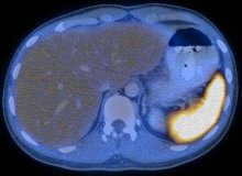
Figure 1.
Reformatted coronal FDG-PET (A) and axial fused FDG-PET/CT (B) images of the abdomen.
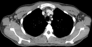
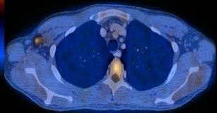
Figure 2.
Axial contrast-enhanced CT (A) and fused FDG-PET/CT (B) images through the chest at the level of the axillae.
Show the video
References
- Kumar V. Abbas A. Fausto N. Robbins and Cotran Pathologic Basis of Disease. 2005. Elselvier. Pennsylvania.
- Nowak M, Carrasquillo JA, Yarboro CH, Bacharach SL, Whatley M, Valencia X, et al. A pilot study of the use of 2-[18F]-fluoro-2-deoxy-D-glucose-positron emission tomography to assess the distribution of activated lymphocytes in patients with systemic lupus erythematosus. Arthritis Rheum. 2004;50(4):1233-8.
- Von Shulthess G. Molecular Anatomic Imaging. 2007.Lippincott Williams & Willkins. Philadelphia.


