Case 20
Dr Marina-Portia Anthony /Dr Henry Mak
Clinical notes
A 70 year-old man presented with 1 day of frank haematuria, on a background of 4 months treatment for polycythemia.Images
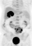
Figure 1.
Whole-body maximum intensity projection (MIP) FDG-PET frontal image.
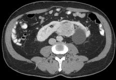
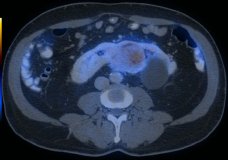
Figure 2.
Axial contrast-enhanced CT (A) and fused FDG-PET/CT (B) images through the mid-abdomen.
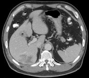
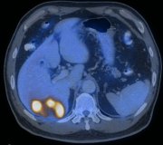
Figure 3.
Axial contrast-enhanced CT (A) and fused FDG-PET/CT (B) images through the liver.
References
- Anthony MP, Mak H, Khong PL. An unusual case of synchronous renal cell carcinoma in a horseshoe kidney and intrahepatic cholangiocarcinoma. Clin Nucl Med 2009;34:922-3.
- Dahnert W. Radiology Review Manual. 2003. Lippincott Williams & Wilkins. Philadelphia.
- Kumar V. Abbas A. Fausto N. Robbins and Cotran Pathologic Basis of Disease. 2005. Elselvier. Pennsylvania.
- Lee C, Hilton S, Russo P. Renal mass within a horseshoe kidney: preoperative evaluation with three-dimensional helical computed tomography. Urology. 2001;57(1):168.
- Rubio B, Regalado P, S?nchez M, et al. Incidence of tumoural pathology in horseshoe kidneys. Eur Urol. 1998;33(2):175-9.
- Tobias-Machado M, Massulo-Aguiar MF, Forseto PH Jr, et al. Laparoscopic left radical nephrectomy and hand-assisted isthmectomy of a horseshoe kidney with renal cell carcinoma. Urol Int. 2006;77(1):94-6.
- Von Shulthess G. Molecular Anatomic Imaging. 2007.Lippincott Williams & Willkins. Philadelphia.


