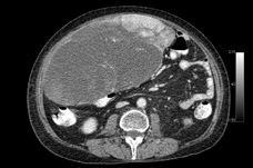
A. Axial contrast-enhanced CT image of the abdomen demonstrates a heterogeneous soft tissue mass in right anterior abdomen extending to the pelvis, approximately 22 x 12 x 25cm in size. The peripheral parts of the mass are solid and hypervascular, as demonstrated by the early enhancement.
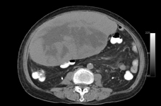
B. Axial delayed contrast-enhanced CT image of the abdomen demonstrates progressive contrast enhancement in the periphery of the mass. The central part of the mass is hypoenhancing and may be necrotic or cystic.
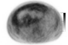
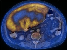
C, D. The peripheral enhancing portion of the mass is mildly hypermetabolic, with SUVmax of 2.2, whereas the central portion is hypometabolic.
E. Large hypermetabolic mass is again noted in the right anterior abdomen.


