Case 19
Dr Marina-Portia Anthony /Dr Henry Mak
Clinical notes
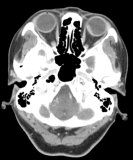
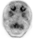
A 68-year-old woman presented with left hip pain for one month, found to be a lytic lesion in the greater trochanter on plain radiograph. Whole body FDG-PET/CT was requested to look for a primary malignancy and other metastases.Images
Figure 1.
Axial images of the lower brain/ skull base on contrast-enhanced CT (A), FDG-PET (B), and FDG-PET/CT (C).
References
- Donaldson MJ, Pulido JS, Mullan BP, et al. Combined positron emission tomography/computed tomography for evaluation of presumed choroidal metastases. Clin Experiment Ophthalmol. 2006;34(9):846-51.
- Nguyen BD, Roarke MC. Choroidal and extraocular muscle metastases from non-small-cell lung carcinoma: F-18 FDG PET/CT imaging. Clin Nucl Med. 2008;33(2):118-21.
- Nguyen NC, Akduman EI, Sayed MH, et al. Increased 18F-FDG uptake in the posterior ocular bulb is associated with brain metastasis: a retrospective study. AJR Am J Roentgenol. 2008;191(6):W268-74.
- Su HT, Chen YM, Perng RP. Symptomatic ocular metastases in lung cancer. Respirology. 2008;13(2):303-5.


