Case 18
Dr Marina-Portia Anthony /Dr Henry Mak
Clinical notes
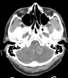
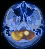
A 56 year-old male presented with headache, nausea and unsteady gait. An MRI showed a left cerebellar tumour with oedema and obstructive hydrocephalus. A PET/CT was requested to determine if the lesion was primary or secondary.Images
Figure 1.
Axial contrast-enhanced CT (A), fused PET/CT (B) images through the cerebellum.
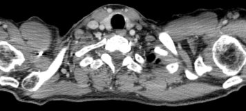
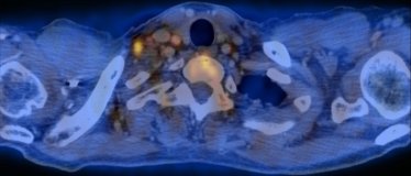
Figure 2.
Axial contrast-enhanced CT (A), fused PET/CT (B) images through the supraclavicular region.
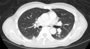
Figure 3.
Axial contrast-enhanced CT (A), fused PET/CT (B) images through the mid chest.
Show the video
References
- Chorost M, Lee C, Yeoh C, et al. Unknown Primary. Review Article. J Surg Oncol 2004;87:191-203.
- Dong M, Zhao K, Lin X, et al. Role of fluorodeoxyglucose-PET versus fluorodeoxyglucose-PET/computed tomography in detection of unknown primary tumor: a meta-analysis of the literature. Nucl Med Commun. 2008;29(9):791-802.
- Kumar V. Abbas A. Fausto N. Robbins and Cotran Pathologic Basis of Disease. 2005. Elselvier. Pennsylvania.
- Kwee T, Kwee R. Combined FDG-PET/CT for the detection of unknown primary tumors: systematic review and meta-analysis. Eur Radiol. 2009;19(3):731-44.


