Case 17
Dr Marina-Portia Anthony /Dr Henry Mak
Clinical notes
A 61 year-old man presented with intermittent fever on a background of lupus nephritis. Septic work-up was negative.Images
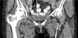
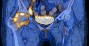
Figure 1.
Reformatted coronal contrast-enhanced CT (A) and fused FDG-PET/CT (B) images through the hips.
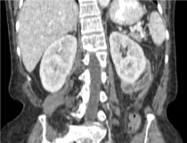
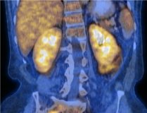
Figure 2.
Reformatted coronal contrast-enhanced CT (A) and fused FDG-PET/CT (B) images through the kidneys.
References
- Von Shulthess G. Molecular Anatomic Imaging. 2007.Lippincott Williams & Willkins. Philadelphia.
- Dumarey N, Egrise D, Blocklet D, Stallenberg B, Remmelink M, del Marmol V, et al. Imaging infection with 18F-FDG-labeled leukocyte PET/CT: initial experience in 21 patients. J Nucl Med. 2006;47(4):625-32.
- Pugh KW, Seligson D, Turbiner E. Positron emission tomography in orthopedics. J Ky Med Assoc. 2004;102(6):259-61.


