Case 1
Dr Marina-Portia Anthony / Dr Alex Ching / Dr Jingbo Zhang
Clinical notes
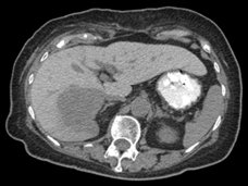
A 71 year-old female presented with an incidental finding of a right hepatic lobe mass, which was hypovascular on contrast-enhanced CT.Images
Figure 1.
Axial non-contrast CT (A) and fused PET/CT (B) images through the upper abdomen.
Show the video
Figure 2.
Cine MIP
References
- Breitenstein S, Apestegui C, Clavien P. Positron emission tomography (PET) for cholangiocarcinoma. HPB (Oxford). 2008;10(2):120-1.
- Kumar V. Abbas A. Fausto N. Robbins and Cotran Pathologic Basis of Disease. 2005. Elselvier. Pennsylvania.
- Petrowsky H, Wildbrett P, Husarik D, et al. Impact of integrated positron emission tomography and computed tomography on staging and management of gallbladder cancer and cholangiocarcinoma. J Hepatol. 2006;45(1):43-50.
- Sun L, Wu H, Guan Y. Positron emission tomography/computer tomography: challenge to conventional imaging modalities in evaluating primary and metastatic liver malignancies. World J Gastroenterol. 2007;13(20):2775-83.


